Cytopathic Effect
Additionally, a total of 1190 photographs were taken as experiment set to gauge the restrict of the mannequin, including 824 from influenza-contaminated MDCK cells and 366 from mock-infected MDCK cells. The picture numbers for the other viruses set had been one hundred twenty. A whole of six non-influenza viruses were included and twenty photos from each virus infected cells had been included for evaluation. The output of different viruses set was adverse. The detail could be revealed within the result section. If the non-influenza virus induced cytopathic effects on MDCK cells, these areas would be chosen for picture taken.
For comparability, remdesivir, the nucleotide analog inhibitor of RNA-dependent RNA polymerase for a variety of viruses and high clinical candidate for SARS-CoV-2 , exhibited an EC50 of seven.04 µM with no obvious cytotoxicity (Fig. 3D). The EC50 values for the entire autophagy inhibitor compounds are summarized in Table 1. Clomipramine was the third finest hit with an EC50 of thirteen.6 µM while inducing less than 20% cytotoxicity at 30.0 µM (Fig. 2B). Hycanthone came in fourth with an EC50 of 5.79 µM and a cytotoxicity CC50 of 14.zero µM (Fig. 2C).
Although not totally absent, apoptotic features were not prominent in a large variety of samples of CD4+ T cells present process death after HIV infection. To quantify these observations, we photographed a variety of sections at low magnification and scored a lot of particular person cells for apoptotic, necrotic, or regular morphology (Fig. 4). These information revealed that a preponderance of apoptotic cells was only seen upon staurosporine remedy in either uninfected or contaminated cells. In contrast, virus infection dramatically elevated the number of necrotic cells but not the variety of apoptotic cells. Notably, staurosporine also considerably elevated the number of necrotic cells in both uninfected and infected cultures.
Autophagy Assays
In panel C, the arrowhead indicates budding virions. The inset in panel C represents a 2.2-fold magnification of the region indicated by the arrow and illustrates the finding of mature retroviral particles inside the particles of a necrotic cell. HIV-1 causes a profound cytopathic effect on cultured CD4+ T lymphocytes from peripheral blood. Purified CD4+ T lymphocytes had been activated with concanavalin A and IL-2 after which infected with the NL4-3HSA strain of HIV-1. Duplicate uninfected or contaminated cultures had been analyzed by circulate cytometry for the fraction of viable cells and the expression of HSA as indicated .
The whole volume in each pattern properly was three ml. After the addition of viral stock, the plates had been centrifuged at 800 × g for 30 min after which incubated at 37°C. Cultures have been maintained by daily cytokine feeding. In experiments with indinavir (IND; AIDS Repository), a ten μM focus was added to the suitable samples previous to centrifugation, after centrifugation, and every day all through the course of the infection. We employed a cell-based mostly assay utilizing Vero-E6 host cells that measures the CPE of SARS-CoV-2 (Fig. 1). The CPE discount assay is a broadly-employed assay format to display screen for antiviral brokers, and it can be scaled for prime-throughput screening .
Viable cells were determined by ahead scatter versus aspect scatter. Infected cells are detected from cells in the viable gate. Note that early HSA is “donated” to the target cells by the virions which have acquired this membrane protein from the producer cells prior to 50 h in this experiment, whereas later HSA is due to provirus expression . For the CPE, inter-plate duplicates had been used for each information level for quantitative HTS and curve fitting.
The experimental definition of viral cytopathicity resulting in the demise of the host cell was established in poliovirus . The remark that poliovirus brought on a severe biochemical derangement of the host cell machinery so that the cell would die offered an important perception into viral pathogenesis. Infected cell death and dysfunction are incessantly related to organ injury and are believed to play an necessary a part of virus-induced illness pathology . Cytopathic results have been noticed for a lot of viruses together with the human immunodeficiency virus .
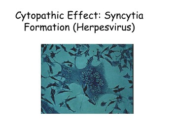
Classical Examples Of The Cytopathic Effect
For a full description, see Giemsa-Stained Bovine Adenovirus -Infected Bovine Fetal Spleen Cells Showing Inclusions. Unstained bovine fetal spleen cells four days postinfection with a excessive MOI of bovine adenovirus, an Adenovirus, exhibiting cell rounding and small amounts of clumping. Unstained bovine fetal spleen cells 2 days postinfection with a excessive MOI of bovine herpesvirus 1, a Herpesvirus. Black arrows level to cell rounding in a focal sample and blue arrows point to cytoplasmic stranding. Yoshida M. Mechanism of trancriptional activation of viral and mobile genes by oncogenic protein of HTLV-1. Infection by cytocidal viruses is usually related to modifications in cell morphology, in cell physiology and sequential biosynthetic occasions.
Kinetic stay-cell imaging of whole-wells, using automated scanning and image stitching software program. Image full properly plates for hours to weeks at a time. CPE is a very primary method to know how a virus infects a cell, however that does not mean it is just used in primary scientific analysis. Measuring CPEs can also be a very useful readout for pharmaceutical firms and diagnostic laboratories. Morphological quantification can be performed utilizing the gating operate in the Celigo software to particularly establish cells of various sizes, smoothness, aspect ratio, mean and integrated intensities. The green outlines determine the counted cells in the properly.
You just want to find the best virus-cell mixture. In the laboratory, a straightforward way of killing a mosquito cell line (like C6/36) or Vero cells is by infecting it with any famous arbovirus, like Chikungunya, Dengue or Zika. After a couple of days of an infection the cells just fall apart proper in entrance of your eyes (Fig. 1).